Home
/ Horse Leg Bones Diagram - 3 _ Finally, there is the large modern horse, equus, with only one toe, while all that is left of the other two are 'vestigial' splint bones.
Horse Leg Bones Diagram - 3 _ Finally, there is the large modern horse, equus, with only one toe, while all that is left of the other two are 'vestigial' splint bones.
Horse Leg Bones Diagram - 3 _ Finally, there is the large modern horse, equus, with only one toe, while all that is left of the other two are 'vestigial' splint bones.. A horse with a broken leg is usually killed because it's very difficult for the broken leg of a horse to heal correctly. The arterial supply to the digit and fetlock of the thoracic limb comes mainly from the median palmar artery.the median palmar artery divides in the distal fourth of the metacarpus between thesuperficial and deep digital flexor tendons and the suspensory ligament, to become the medial and lateral digital arteries.part of the deep palmar arch anastamoses with the lateral digital. Horse leg structure there are also many fossil remains of horse leg bones. The ideal horse has legs which are straight, correctly set and symmetrical. This diagram shows the superficial layer of the tissue.
The pedal bone itself has an unusually high density of blood vessels within it. Hoof and lower leg structure. The limbs of the horse are structures made of many bones, joints, muscles, tendons and ligaments that support the weight of the horse's body. One bone works in relation to another. This is because there are many layers of muscles.
Horse Hoof Anatomy Inside And Out from www.equinespot.com Finally, there is the large modern horse, equus, with only one toe, while all that is left of the other two are 'vestigial' splint bones. Human anatomy muscles coloring pages printable via. This is logical, as a horse moves forward the bones of the lower leg shield the soft tissues. The shoulder joint is the articulation between the glenoid cavity of the scapula and the head of the humerus.in the horse, lateral and medial movements of this joint are impossible due to the shape of the humeral head; This is because there are many layers of muscles. One bone works in relation to another. Which is the oldest horse on the diagram? Diagram of leg bones and joints via.
Degenerative disease, similar to arthritis.
The horse leg anatomy in the rear includes the bones of the pelvis (the ilium, ischium and pubic bones), femur, tibia, fibula, metatarsus and the phalanxes. We make sure to keep the original photos without changing. In our website, we are persons who very treasure creativity from every one, with no exception. Horse leg bones diagram quizlet. The limbs play a major role in the movement of the horse, with the legs performing the functions of absorbing impact, bearing weight and providing thrust. They are joined to the spine through the sacroileac joints and allow transfer of propulsion to the hind legs. Which is the oldest horse on the diagram? Whereas, the deep digital flexor tendon runs down the back of the leg and wraps around the navicular bone, bending and flexing the leg. Cow horse leg foot muscle and skeleton anatomy. Talking about horse anatomy worksheets printable, we have collected some related images to complete your references. For example, the body part that is called a horses 'knee' is actually the carpal bones that correspond to the human wrist. The horses legs and hooves are also unique, interesting structures. Horse body parts diagram, horse skeleton diagram and animal nervous system diagram are some main things we want to present to you based on the gallery title.
The shoulder joint is the articulation between the glenoid cavity of the scapula and the head of the humerus.in the horse, lateral and medial movements of this joint are impossible due to the shape of the humeral head; This system works together to support horses weight when it stands up also works to diminish compression during movement which helps to horse to avoid injury to their limbs. The power propulsion system and major defensive tool, a horse's rear. The joint is strengthened by the medial and lateral glenohumeral ligaments. The small bone that forms the point of the hock is actually similar to the human heel bone.
Horse Foot Anatomy The Equinest from www.theequinest.com The horse leg anatomy in the rear includes the bones of the pelvis (the ilium, ischium and pubic bones), femur, tibia, fibula, metatarsus and the phalanxes. How many front toes did the oldest horse have? Finally, there is the large modern horse, equus, with only one toe, while all that is left of the other two are 'vestigial' splint bones. This is because there are many layers of muscles. Degenerative disease, similar to arthritis. In this picture it shows the muscles that are closest to the surface of the skin, making them superficial. Cow horse leg foot muscle and skeleton anatomy. They are joined to the spine through the sacroileac joints and allow transfer of propulsion to the hind legs.
Look at the diagram on the previous page of the front legs and toes (hooves) of some of these horse fossils.
This is because there are many layers of muscles. The hoof is heavily supplied with blood through the two arteries which run down the back of the leg and into the foot. Principles of bone development in horses. Lower extremity arterial duplex via. Finally, there is the large modern horse, equus, with only one toe, while all that is left of the other two are 'vestigial' splint bones. The horse leg anatomy in the rear includes the bones of the pelvis (the ilium, ischium and pubic bones), femur, tibia, fibula, metatarsus and the phalanxes. Horse body parts diagram, horse skeleton diagram and animal nervous system diagram are some main things we want to present to you based on the gallery title. The small bone that forms the point of the hock is actually similar to the human heel bone. Cow horse leg foot muscle and skeleton anatomy. The joint is strengthened by the medial and lateral glenohumeral ligaments. The ischium forms the point of the buttock. This system works together to support horses weight when it stands up also works to diminish compression during movement which helps to horse to avoid injury to their limbs. The shoulder joint is the articulation between the glenoid cavity of the scapula and the head of the humerus.in the horse, lateral and medial movements of this joint are impossible due to the shape of the humeral head;
There are many possible diagrams of the anatomy of horse tissues. Individual horses may have structural defects, some of which lead to poor. This is supposed to demonstrate a change from browsing on bushes to grazing on grass. The photograph shows the laminae which keep the hoof wall tightly bonded to the internal structures. The shoulder joint is the articulation between the glenoid cavity of the scapula and the head of the humerus.in the horse, lateral and medial movements of this joint are impossible due to the shape of the humeral head;
How Equine Forelimb Anatomy Plays Out With Conformation And Soundness from www.equinespot.com Start studying front horse leg. The legs of a horse are made up of a system of muscles, tendons, ligaments, and connective tissue. The bulk of soft tissue is behind the bones. Lower extremity arterial duplex via. The limbs play a major role in the movement of the horse, with the legs performing the functions of absorbing impact, bearing weight and providing thrust. If the angle at witch these bones are working is compromised, the joint becomes unevenly stressed and injury to the tendons and ligaments. Hoof and lower leg structure. This is because there are many layers of muscles.
When the skeletal structure is properly proportioned the joints work smoothly.
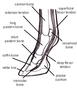
The eohippus horses part b. The power propulsion system and major defensive tool, a horse's rear. Human anatomy muscles coloring pages printable via. The top part of the hind limbs consists of three fused bones, called the ileum, ischium, and pubis. For example, the body part that is called a horses 'knee' is actually the carpal bones that correspond to the human wrist. Inflammation of navicular bone and/or bursa. The bulk of soft tissue is behind the bones. The bones that make up the lower leg are the cannon bone, splint bones, long pastern, short pastern, pedal bone and navicular bone. Similarly, the hock contains the bones equivalent to those in the human ankle and heel. Individual horses may have structural defects, some of which lead to poor. See more ideas about horse. Movement is therefore limited to flexion and extension. The ideal horse has legs which are straight, correctly set and symmetrical.
In the front of the leg, the only thing covering the bones is skin, the extensor tendons (which are very flat) and the suspensory ligaments and fascia leg bones diagram. Talking about horse anatomy worksheets printable, we have collected some related images to complete your references.

